
In the past ~15,000 years, humans have undergone a collapse in form. This likely includes yourself and those around you
Whole Body Breathing theory and practice aims to understand and correct the physical, emotional, and esoteric distortions in humans.
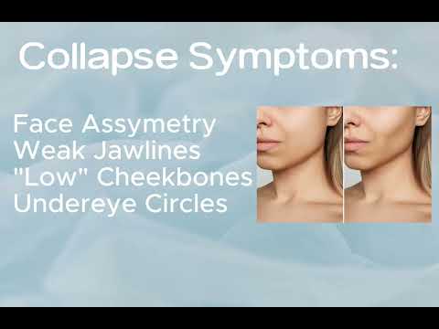
The following video is a general overview of the symptoms of collapse and results from Whole Body Breathing:
Section 0: Information and Overview
[VIDEO OVERVIEW HERE]
This /learn page will link you to every possible part of the project. There are group classes, written articles, social media channels, group chats, newsletters, practitioners, devices etc - the scope of the project is huge. I suggest that you click around to get your bearings and then dive in wherever seems logical/calls to you.
Outline of the Learn Page
Section 0 Information and Overview
You are here
Section 1 Collapse and Correction
1.1 Symptoms and Causes
1.2 Correction Methods
Section 2 Whole Body Breathing
2.1 Introduction to Whole Body Breathing Theory and Routines
2.1 Psychosomatics, Chemistry, and Energy Correction Theory
2.1.1 Exercises, Techniques, Devices
2.3 Physical Structure Correction Theory
2.3.1 Exercises, Techniques, Devices
Section 3 Project Praxis
Case Discussions
Testimonials
Become a Practitioner / Sponsor
Activism Campaigns
Project Roadmap
Required Research
Public Group Classes + Group Chats
Group Classes are held sporadically. Use the group chats below to get notifications (You can join one or many).
Add your best times here
Add your best times here
Join the Discord Announcements Server (Click here)
You do not need to finish the sections/modules before joining the classes
Some classes are myself teaching discussion topics, ending with QA and Meditations. Other Classes are QA and Meditation only.
Practitioners
Social Media Channels
Book
The Book is Under Development
Podcast
The Podcast is Under Development
Section 1: Collapse and Correction
The physical, mental and spiritual form of humans has collapsed in the past 15,000 years.
Symptoms and CausesThe Symptoms and Causes page discusses the extent of collapse - the widespread potential symptoms and the myriad of interlinked potential causes. There are further Discussions inside the Symptoms and Causes Page.
The Correction Methods page dives into the many methods which have been developed for correcting the collapse. Some methods are aware of the collapse and try to address its totality, while others deal with individual aspects. Some methods are new while others are very ancient. The relationship of these methods with WBB is explored. There are further Discussions inside the Corrections Methoid Page.
Section 2: Whole Body Breathing
Whole Body Breathing is an advanced method of breath awareness which unlocks your proprioception of the pressurized fascial tensegrity system which makes up your body, including 7 diaphragms and 2 main fascial tension lines connecting them. Through this increased awareness, improper tension is released and proper tensioning can be rebuilt.
This is a psychosomatic and yogic experience, as the body and mind are not separate, and many of the tension patterns in the body are reflections of egoic tension and trauma known as Samkoca.
Attending live classes is highly recommended. Click here to see how
Section 3: Project Praxis
This section is for the development of a movement which will bring the collapse/treatment discussion into the mainstream, develop awareness of practices which are harmful to development, creating/funding research into causes and treatments (and of a correct working model of the body/posture/breathing/movement), organizing groups and campaigns etc. If you would like to get involved in the project, this is certainly the place to do so.